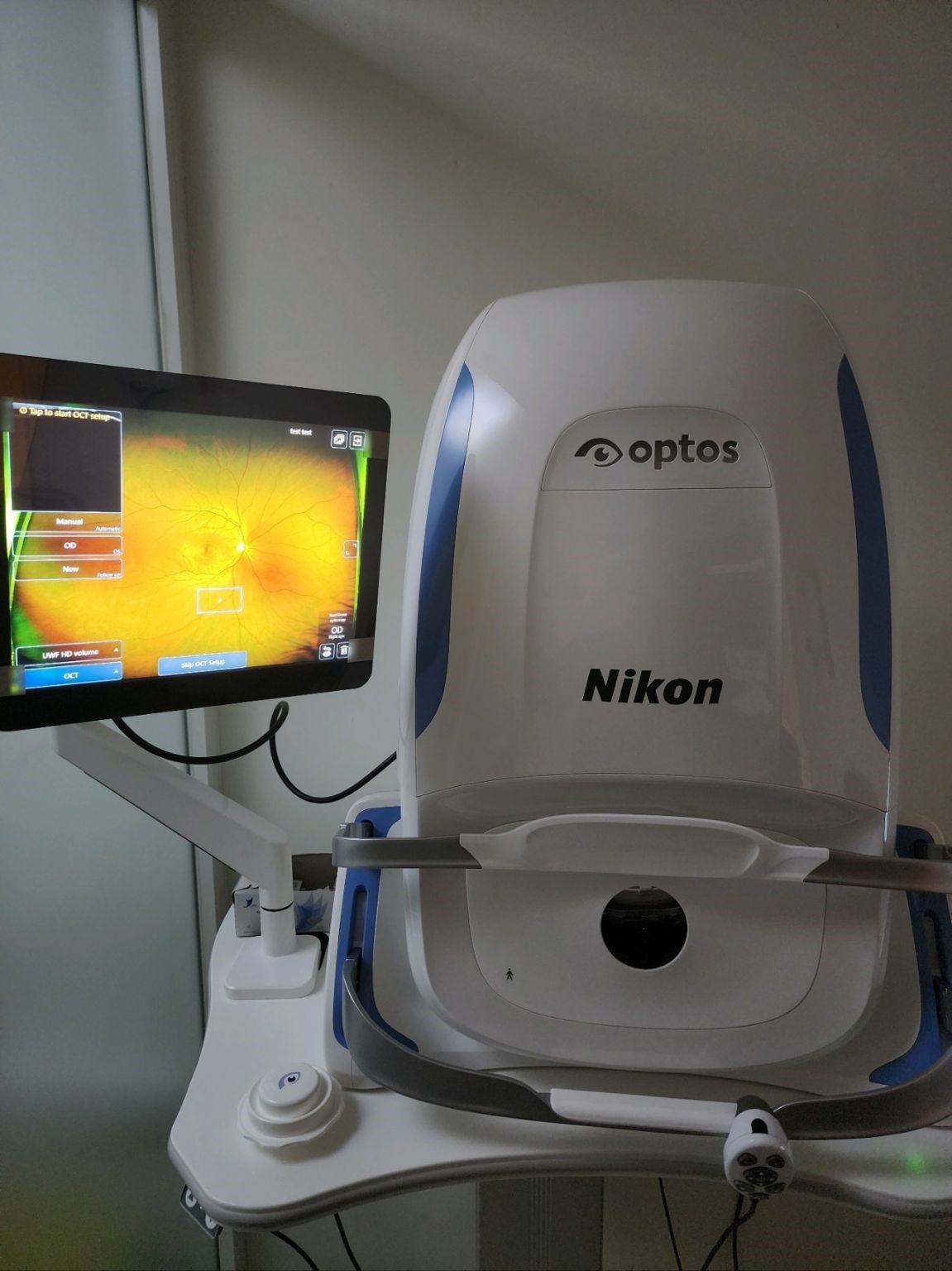
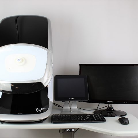
SILVERSTONE RGB
Optos Silverstone RGB Retinal Imaging platform, and delivers a suite of 9 imaging modalities in a single device. Silverstone RGB offers a broad spectrum of retinal imaging capabilities, enabling clinicians to capture, visualize, and analyze pathology across every layer of the retina.
9 modalities available with the Silverstone RGB device include: optomap color RGB for true-to-life retinal color imaging, optomap color RG, optomap fluorescein angiography for vascular evaluation and disease detection, optomap indocyanine green angiography for enhanced choroidal and vascular imaging, optomap sensory red-free for highlighting nerve fiber and vascular structures, optomap choroidal imaging for deep tissue visualization, optomap green autofluorescence for identifying RPE changes and metabolic activity, optomap blue autofluorescence for visualizing subtle retinal pathology, and swept-source OCT for high-resolution, navigable imaging anywhere in the retina.
Benefits
Nine imaging modalities and UWF with integrated Swept Source OCT, facilitate detailed examination of the retina-vitreous to sclera
UWF guided, Swept Source OCT, images pathology anywhere on the optomap
1050 nm OCT light source, provides deeper tissue penetration for clear, detailed choroidal imaging
4-in-1 Color Depth ImagingTM provides important clinical data from the retinal surface through the choroid
Image Modalities
• Optomap color rgb (red, green and blue laser)
• Optomap color rg (red and green laser)
• Optomap Sensory Retina (green laser)
• Optomap Choroidal (red laser)
• Optomap green af (green laser): autofluorescence
• Optomap blue af (blue laser): autofluorescence
• Optomap fa (blue laser): fluorescein angiography
• Optomap icg (infra-red): indocyanine green angiography
• Swept Source Optical Coherence Tomography (OCT)
Resolution
• Optomap plus: 14 μm
• Optomap: 20 μm
Wavelengths
• Red laser: 635 nm
• Green laser: 532 nm (for af )
• Blue laser: 488 nm (for fa)
• Infra-red: 802 nm (for icg)
OCT Imaging
• Signal Type: Optical scattering from tissue
• Signal Source: Swept source OCT, Wavelength 1050 nm
• Optical Power: Laser safety Class-1 following IEC/en60825-1:2014(2007)
• Axial Resolution: <7 micron
• Transverse Resolution: <20 micron (in tissue)
• Scanners: Galavanometric with x, y pair
• Scan Depth: Up to 2.5mm
There are no reviews yet.