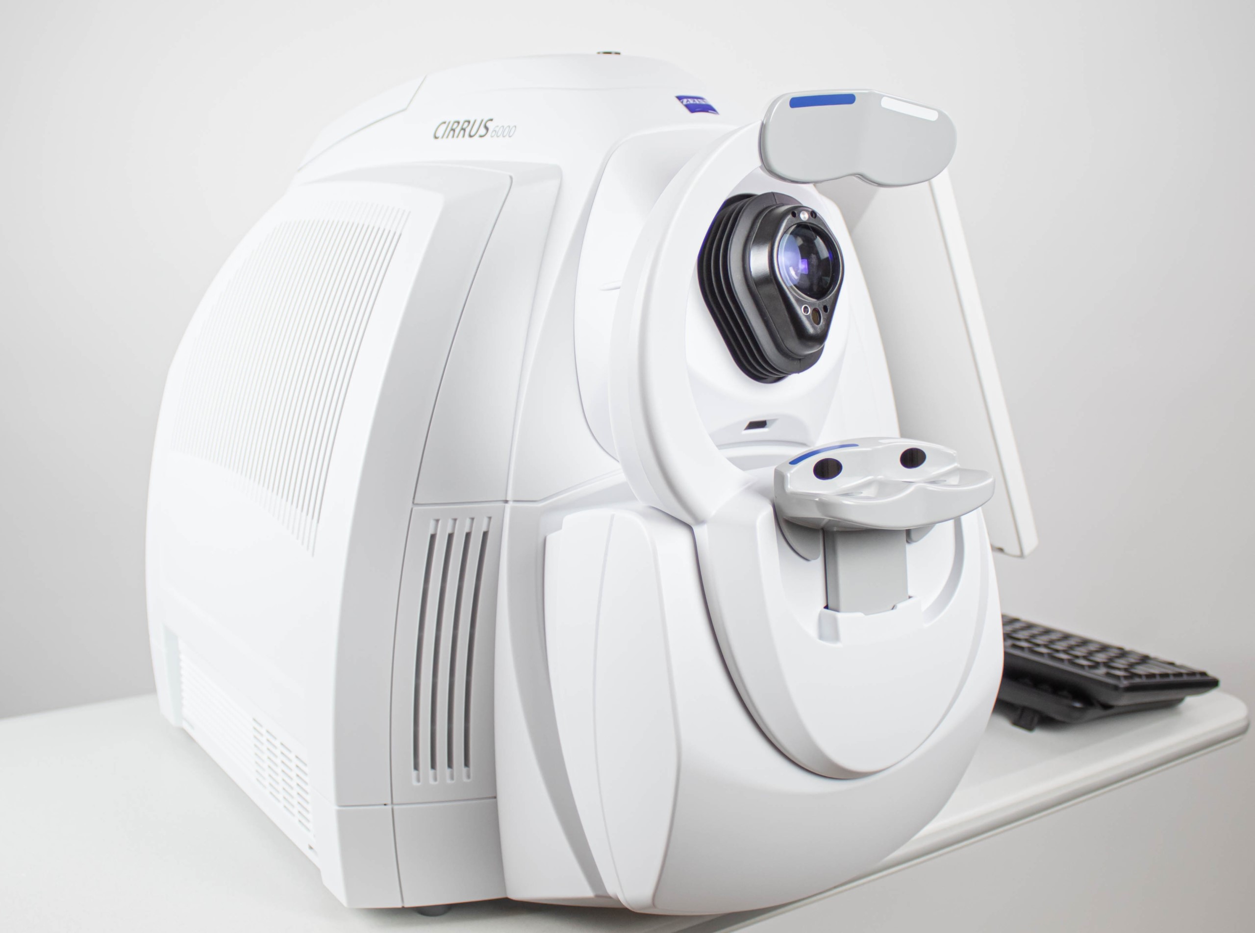
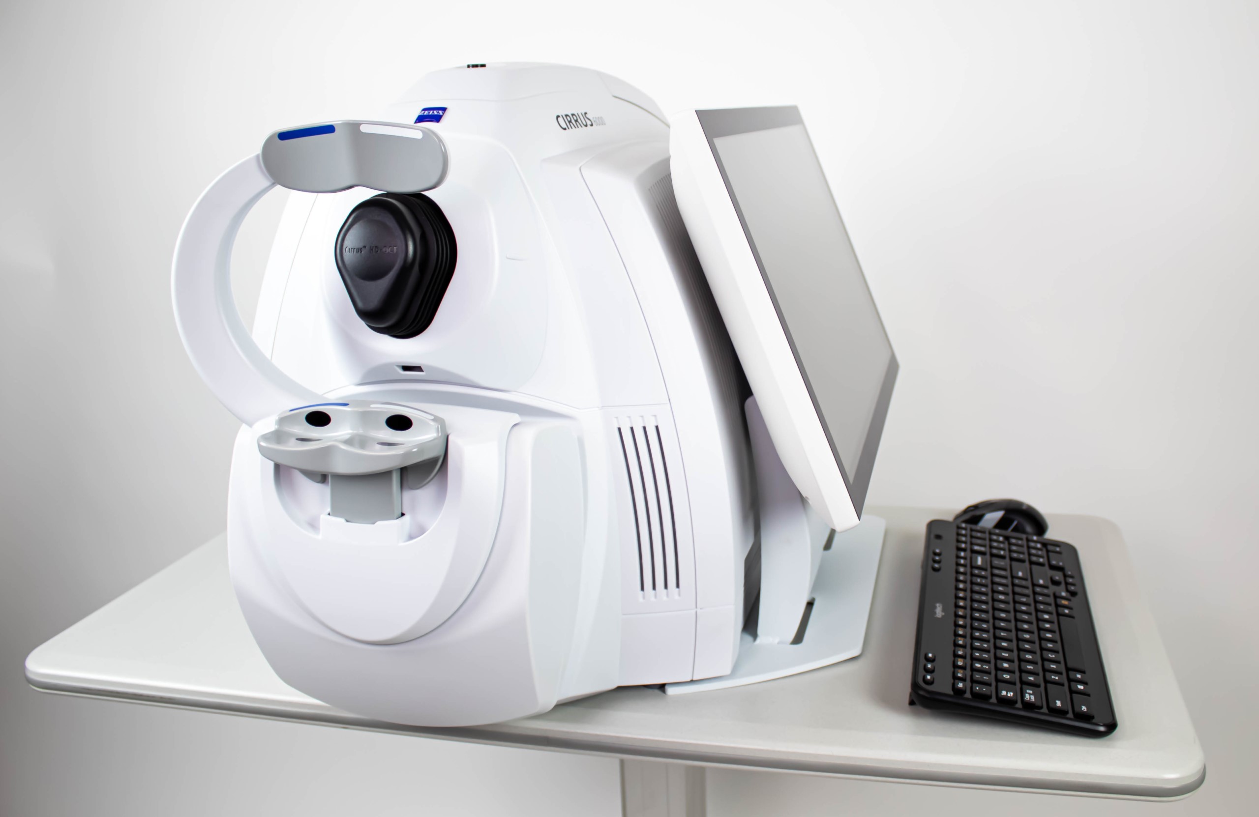
CIRRUS 6000
High-Performance OCT Advance your fast-paced practice CIRRUS 6000, the next-generation OCT from ZEISS, delivers high-speed image capture with HD imaging detail and a wider field of view, so you can make more informed decisions and spend more time with your patients. CIRRUS 6000 is the latest addition to an already robust portfolio of OCT devices that are designed to meet the needs of all types and levels of practices and/or clinicians. It offers customers high-speed and high-throughput to help manage their large patient volume faster, all while improving imaging and image quality.
Performance OCT: Faster imaging with greater detail, at 100,000 scans per second, for improved patient care.
Proven analytics: Comprehensive, clinically validated tools to diagnose and manage a range of conditions.
Patient-first design: Seamless transfer of raw patient data from previous generations of CIRRUS for continuity of patient care.
The power of 100,000 scans per second
Faster imaging: Reduce chair time and speed up your practice.
- 270% faster OCT scans and 43% faster OCTA scans
- OCT cube scans in as little as 0.4 seconds
- High-speed imaging in combination with FastTrac eye tracking technology reduces the chance of motion artifacts such as those caused by blinks and saccades
Greater detail: View more in seconds and dig deeper with high-definition imaging.
- 12×12 mm single-shot OCTA cube scan in addition to 8×8, 6×6 and 3×3 mm scans
- High-Definition AngioPlex scans (8×8 and 6×6 mm) for even greater microvascular
- 2.9 mm scan depth
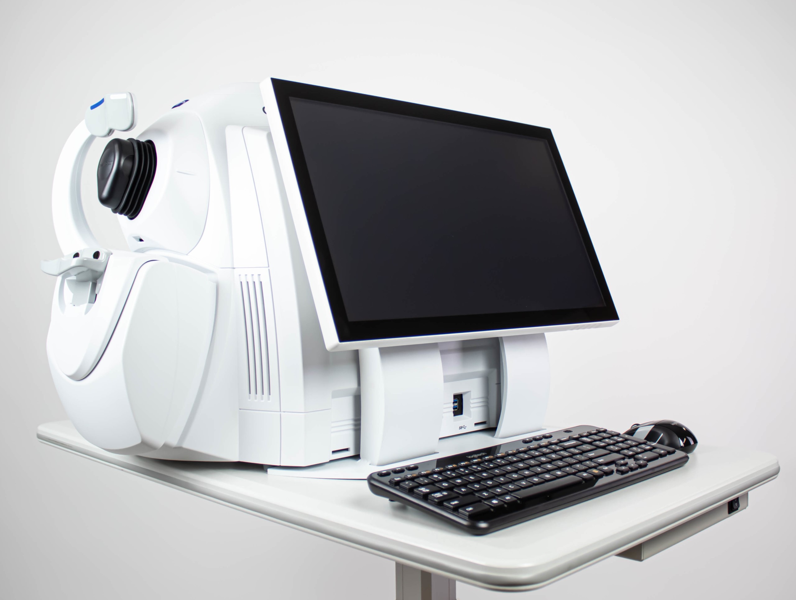
Performance OCT — faster, wider, with a new level of detail
ZEISS CIRRUS 6000 empowers clinicians with a larger field of view in a single scan, and captures high-definition OCT/OCTA scans that reveal finer details of the retinal microvasculature − all of which provides more insight into the patient’s condition in less time.
Proven analytics: CIRRUS-powered treatment decisions
As the pioneering OCT technology, the CIRRUS platform offers clinicians extensive, clinically-validated applications for retina, glaucoma and anterior segment. The result: precise analysis, faster throughput and smarter decision-making across a wide spectrum of clinical conditions and patient types.
Retina
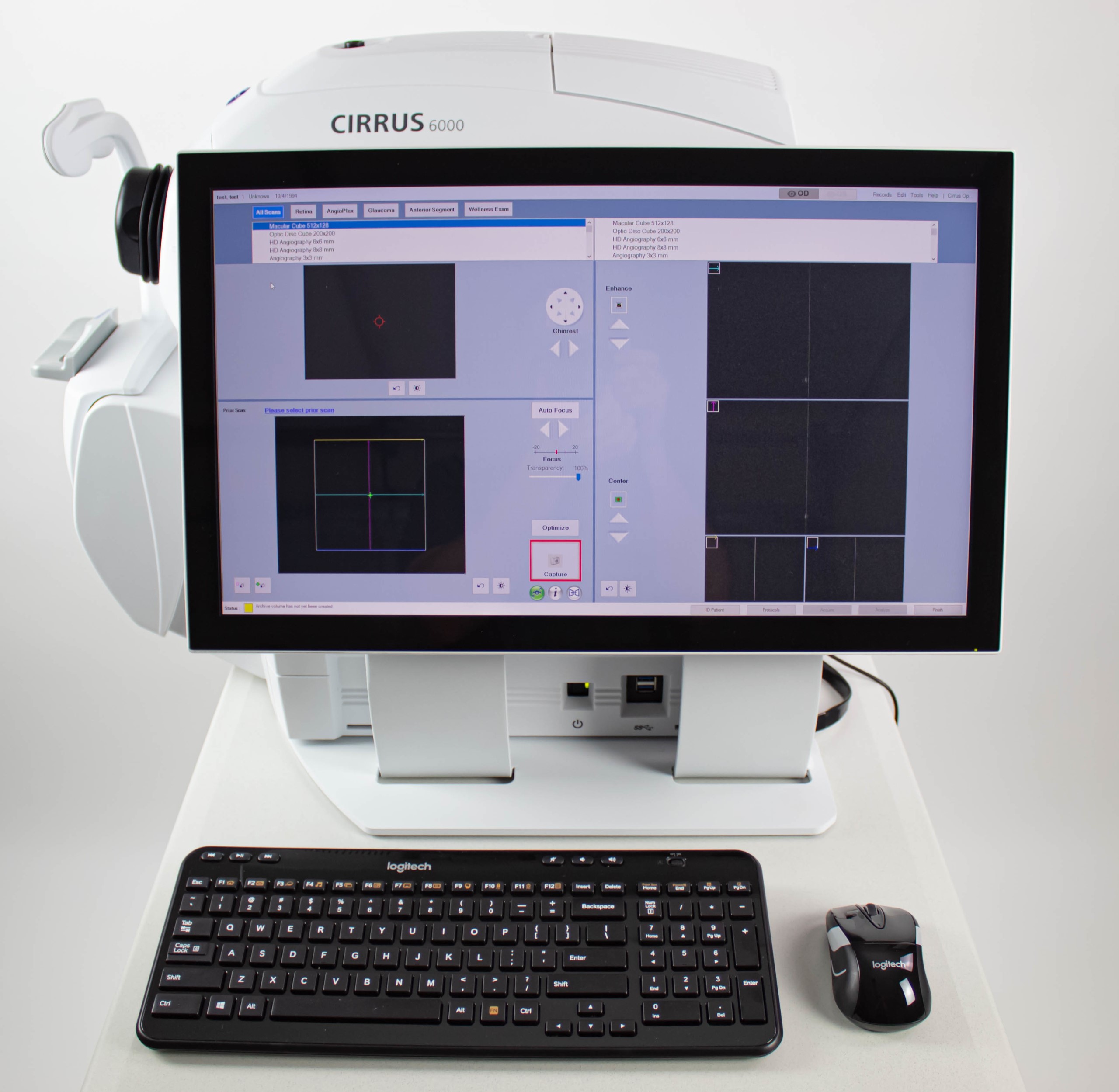
Macular Change Analysis The CIRRUS data cube automatically stores and delivers each patient’s historical data to provide a variety of change assessments, including macular thickness change maps that help you understand your patient‘s response to treatment. Because every CIRRUS cube is tracked and registered to OCT scans from prior visits using CIRRUS’ FastTrac™ Retinal Tracking Technology, you can confidently measure point-to-point changes in macular thickness.
AngioPlex Metrix OCTA Quantification
AngioPlex® Metrix™ for Macula and ONH: AngioPlex Metrix allows clinicians to objectively assess and track progressive eye diseases such as diabetic retinopathy and glaucoma with quantification tools such as Vessel Density, Perfusion Density, and Foveal Avascular Zone (FAZ) for the macula, and Capillary Flux Index for the optic nerve head. *Example In Pictures*
Technical specifications
Key Parameters
- Methodology: Spectral domain OCT
- Optical source: Superluminescent diode (SLD), 840 nm
- A-scan depth: 2.0 – 2.9 mm (in tissue)
- Scan speed: 100,000 A-scans per second
- Min. pupil diameter: 2.0 mm
- Resolution:
• Axial resolution
• Transverse resolution
5 μm (in tissue), 1.95 μm (digital)
15 μm (in tissue) - Refractive error adjustment: -20D to +20D (dopters)
- Resolution:
• Axial resolution
• Transverse resolution
5 μm (in tissue), 1.95 μm (digital)
15 μm (in tissue) - Posterior Segment scans:
• OCT
• OCTA
Cube scan (Macula and Optic Disc)
HD Raster (1, 5, 21-line, cross and radial); Raster scan length 3-12 mm;
image averaging up to 100x
3×3, 6×6, 8×8, 12×12 mm (Macula); 4.5×4.5 mm (Optic Nerve Head);
14×10 mm (Montage), 14×14 mm (Montage)
Analytical applications
Retina:
- Macular Thickness Analysis with Reference Database (Diversified and Asian)
- Macular Change Analysis
- Advanced RPE Analysis
- 3D Visualization
- En Face Analysis
- CIRRUS Wellness Exam
Glaucoma:
- Guided Progression Analysis
- Ganglion Cell/IPL Thickness with Reference Database (Diversified and Asian)
- ONH Parameters with Reference Database (Diversified and Asian)
- Average cup-to-disc ratio
- Average, Superior and Inferior RNFL Thickness
- CIRRUS Wellness Exam
Anterior Segment:
- 9 mm Epithelial Thickness and Pachymetry Mapping
- HD Cornea with Cornea Caliper Tool
- ChamberView™ Full Anterior Chamber Imaging for phakic IOL sizing and safety distance measurements
- Angle imaging and measurement tools for Glaucoma (AOD, TISA, SSA)
AngioPlex Metrix OCT Angiography Quantification:
- Macular
- Foveal Avascular Zone
- Vessel Density (ETDRS grid)
- Optic Nerve Head
- Capillary Perfusion Density
- Capillary Flux Index
- AngioPlex 2-visit comparison
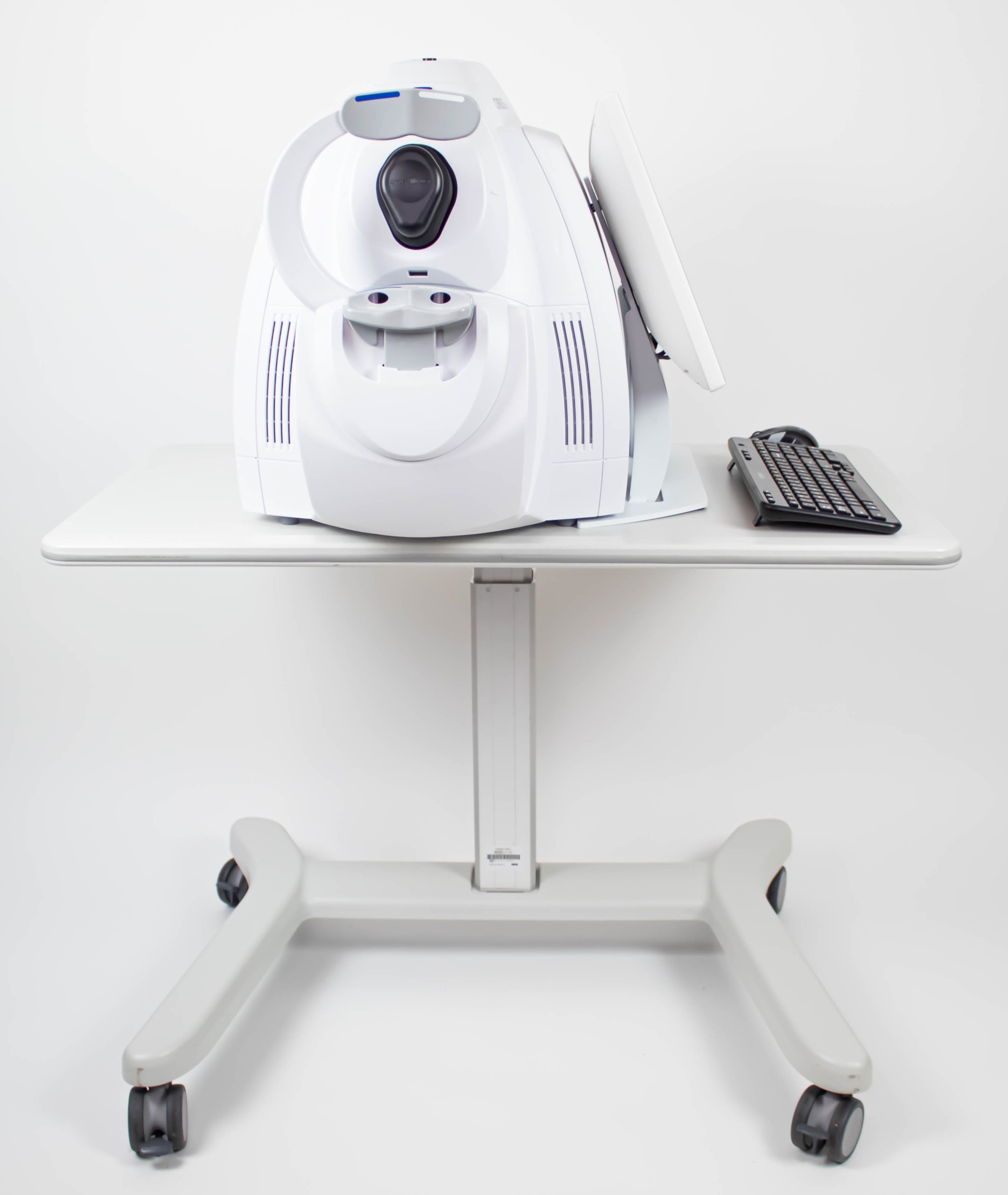
Instrument Specifications:
- Weight: (77 lbs)
- Dimensions (L × W × H): (24.4 × 16.7 × 11.4 in)
This Zeiss Cirrus 6000 comes equipped with:
- 6 Month warranty
- Anterior Segment software
- Angioplex Software
- Power table
There are no reviews yet.